Simulation Modalities Available
Standardized Patients
Part-task Physical Trainers
For many purposes, especially for learning particular tasks and skills, it is only necessary to replicate specific portions of the patient or task. Part-task physical trainers provide just the key elements of the procedure or skill being learned. While they cannot fully replicate performing the task on real patients, they do allow learners to acquire the basic skills needed to then be taught the finer points of doing the procedures under supervision on actual human beings.
Many kinds of part task trainers are used. In some cases, basic elements of a task can first be practiced on household objects or food. Many a student has first learned how to do an injection by practicing on an orange, and residents in anesthesiology may learn to do epidurals (e.g. for relief of pain during labor and childbirth) using a watermelon! For other procedures, more advanced physical trainers that better replicate aspects of human beings are needed. Hybrid programs that combine a part-task physical trainer with a live standardized patient actor are also becoming increasingly common. For example, part-task trainers for suturing of deep cuts or lacerations consist of a pad of a special soft plastic material with a cut in the surface. Such a pad can be strapped on the arm of an SP actor, and then draped with appropriate clinical draping material. The student not only has to perform the elements of sewing up the wound, but, as in real life, has to do so while conversing with the patient and attending to his/her needs.
We have a variety of part-task trainers at the ILC that can be incorporated in to different curricula. You can reserve these resources through Medscheduler.
Virtual Reality and Visualization
Virtual reality refers to a set of techniques in which one interacts with a synthetic (“virtual”) environment that exists solely in the computer. In the typical conception of virtual reality, the representation of the synthetic environment is fed fairly directly to the eyes, ears , and possibly hands. The actions of the user in the environment are translated directly from typical physical activities. There is a continuum of realizations of this ideal, involving compromises on these input/output modalities. At one end of the continuum, which we call a “complete virtual reality simulation,” the participant is immersed in a virtual world that fully replicates at least three sensory inputs—vision, hearing, and touch (the last is more technically known as a haptic/kinesthetic system)—and allows complete physical interaction with the world. At the other extreme of the continuum is a screen-based simulator, which generates a limited virtual world, but it restricts its output to a screen display and provides interaction with the virtual world only through a pointing device. The screen-based simulator provides an interface to the human sensory system that is very far from physical reality, whereas a complete virtual reality simulation may be, in its most advanced form, nearly indistinguishable from the real world. A “partial” virtual reality simulator would replicate fewer senses (or less complete replication, such as a 3D visual representation on a 2D screen) and/or could restrict physical interaction with the world. Finally, one can imagine hybrids of realistic simulators and virtual reality simulators in which the virtual reality representation is overlaid onto a real physical environment.
A variant of virtual reality is often called “visualization”. This involves presentation of 3D structures (such as anatomy or molecular structure) in ways that maximize learning. This can involve true stereoscopic viewing of such structures using special projectors and glasses or special head-mounted displays with a separate view for each eye. Other forms of visualization present only a perspective image on a two-dimensional screen, but with such high resolution and with such extensive means of manipulating the images (such as rotating it, zooming, etc.) that any part can be viewed in detail. Visualization is not only a simulation technique for learning, but also can be used as part of patient care (to plan complex procedures) and to view research data (turning numbers into pictures).
Virtual reality is often used to create advanced part-task trainers for complex medical procedures such as minimally-invasive surgery (e.g. laparoscopic surgery) or catheter-based “endovascular” procedures such as coronary angiography, angioplasty, and stent placement. Virtual reality lends itself to these procedures because the clinician in the real world already is interacting with an image on a screen (from a camera in laparoscopy; from the X-ray fluoroscopy in the “cath-lab”) using artificial extensions of the hands (laparoscopic surgical instruments and long, thin catheters in the blood vessels in the two examples).
Creating a complete virtual reality replication of the entire patient and treatment setting is very complex as it would require the following:
- A complete computer model of the patient, the environment, and the function of every object in the environment that could be utilized (e.g., monitoring devices, carts).
- A means of tracking visual, audio, and touch fields of the user to determine what is to be displayed and to identify what physical actions are being performed.
- Appropriate display hardware for every sensory modality and appropriate input hardware for each action pathway (e.g., touch, speech).
- Hardware to compute all the models, to conduct the tracking, and to produce all the outputs to the display hardware in real time.
Virtual reality is a rapidly developing field. There is intense interest in virtual reality in a number of domains, particularly the military and entertainment. Although the potential of this approach is very exciting virtual
Desktop Simulations and Virtual Worlds
Some kinds of simulations do not need fancy visualization or interaction technologies. They can run on a computer using only a screen, a pointing device, and sometimes audio inputs and outputs. A desktop simulator puts all or part of the patient “in the computer.” One can see the patient via animation, drawings, or video. The learner can interact with the patient by asking questions (typing or speaking) or making comments, by viewing data from monitors, labs, or x-rays, and by performing diagnostic or therapeutic actions (typically by making choices with the mouse). Unlike a standardized patient actor, the simulated patient in a desktop simulator doesn’t mind having a serious life-threatening condition, being stuck with needles or, should it come to that, dying. The simulated patient can respond to whatever the course of the disease is based on the actions taken.
An even more advanced variant of this approach allows multiple participants to interact simultaneously with the same patient in a common “virtual world,” perhaps linked through the Internet from many different site. The virtual world is typically presented as a 2D perspective world. Each participant has an “avatar” that represents them in the virtual world. You can move your avatar, and when you speak, it speaks. Your avatar can interact with the other participants, who may take on different clinical roles in the clinical world that is created. As in the desktop simulation, information can be gathered, actions taken, and the patient’s condition may improve or worsen. The unique aspect of the virtual world is that it directly supports the kinds of interpersonal interactions between various clinicians and with the patient.
Mannequin-based Patient Simulation
Taking the underlying concepts described for the desktop simulation one step further is the recreation of the real physical patient in a realistic physical clinical environment. This is done because real patients don’t live in a virtual world – they and the clinicians who care for them work in an actual physical world with real people. Thus, computerized mannequin stands in for the patient, and a variety of equipment can be used (either real clinical equipment or computer-driven replicas) to monitor and treat the patient.
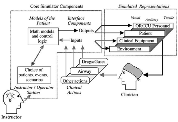
The mannequin-based simulator has a computer representation of the patient similar to that in a desktop simulator, replacing the videos, drawings, and animations with actual functions of the “plastic person.” Among the functions that these mannequin-based simulators can replicate are:
- Spontaneous breathing (and the ability to breathe for the patient with a bag or ventilator)
- Real-time display of electronically monitored information (e.g. ECG, oxygen saturation, etc.)
- Pulses, heart sounds, breath sounds, pupil size, pupil response to light
- Obstruction of various parts of the airway
Not only are these created in their “normal” manifestation, but all the elements of a large variety of abnormal conditions can be created such as (the list is almost endless) heart attack, severe allergic reaction, breathing difficulties, sepsis (“blood poisoning”), severe abnormalities of sugar metabolism, etc.
Some simulators contain complex models of the effects that many different drugs will have, tracking the distribution of the drug in the body and calculating the particular effect that a specific amount of drug will have on the different bodily functions. Most of these simulators provide the ability to stick tubes and needles into various places and to perform other invasive maneuvers such as “shocking the heart” (defibrillation) or applying an external pacemaker.
Mannequin-based simulators typically include speakers in or near the patient’s head so that the mannequin is also a Standardized Patient actor. The voice of the patient can come from the computer, or from an instructor or actor using a microphone. Having an SP that can get seriously ill or die, without complaining, is a real advantage in some settings.
Mannequin-based simulators have become very common in many fields (operating room, intensive care unit, emergency department, labor and delivery rooms) where life-threatening situations require prompt recognition and treatment by individuals or teams of clinicians. Simulations can be conducted in a dedicated simulation learning facility, or portable simulated patients can be taken to empty clinical spots in a real work unit (say a real ICU) where simulation exercises can be conducted in the actual places that patient care is done. These simulators are used across all levels of learners, from K-12 students to early learners in university or medical or nursing school, and by experienced personnel both as individuals, groups, and full multidisciplinary teams. Not only are lessons learned by and about the people in the simulations, lessons are also learned about clinical work units and “systems”. These lessons can be turned into effective improvements in the workplace and beneficial changes in how we take care of patients in the future.
Giving to CISL
Please consider making a gift to CISL.